The Mesh That Makes You Believe: Precision Sieves and the Psychology of Reliable Microplastic Science
Aug 09, 2026
The Biologist’s Dilemma
She stared at the fluorescence microscope image on the screen. The green dots were supposed to be microplastics ingested by the tissue sample. But the background was a hazy cloud of noise. “Is that plastic, or just undigested protein glowing?” she muttered. The data was unusable.
This is the quiet agony of microplastic research. You spend days digesting liver or mussel flesh in caustic chemicals, only to find that what you’ve isolated is a chemical ghost—contaminated, poorly sized, and analytically meaningless. The root of the problem is not the microscope or the spectrometer. It’s something far simpler: a failure of physical separation.
We tend to trust complex machines. We believe in the mass spectrometer, the Raman microscope, the algorithms. But reliable science often rests on a much older, almost forgotten piece of engineering: the precision sieve. A mesh of woven wire, when used correctly, becomes the arbiter of truth.
The Physical Gateway: Solid-Liquid Separation That Defines Your Sample
After the aggressive digestion of biological tissue—using potassium hydroxide, enzymes, or acids—you are left with a murky soup. Suspended in that liquid are your target microplastics, but also a universe of degraded organic sludge.
The Mesh as Interceptor
You pour the digestate onto a precision test sieve, typically with a 100 µm stainless steel mesh. The liquid and dissolved fats pass through. The plastic particles don’t.
This is brute-force physics, not complicated chemistry. Woven wire acts as a size-exclusion barrier. It doesn’t care about chemical potential or surface charges; it simply catches anything larger than its calibrated openings. In a workflow plagued by variables, this mechanical certainty is a psychological relief.
Washing Away Doubt
Once the particles are trapped, the sieve becomes a basin for rinsing. You pour distilled water, then maybe ethanol, over the retained material. The rinsing washes away residual digestive chemicals and fine organics.
Psychologically, you’ve just achieved something profound: you have seen the sample getting cleaner. The turbid filtrate runs clear. You now have a solid residue you can trust. Without this step, the downstream analysis is a bet against background noise—a bet you usually lose.
Standardizing Size: Why a Micron of Variance Breeds Chaos
Imagine you are conducting a toxicological feeding study. Fish A ingests a jagged, 200 µm fiber. Fish B ingests a smooth, 20 µm sphere. Their inflammatory responses will be wildly different. If you can’t control particle size, you can’t interpret your data.
Eliminating the “Dust” Illusion
A single sample contains a mess of particles: fragments, spheres, fibers, and fine dust. Without sieving, your “microplastic” sample includes particles too large to be biologically relevant and fines that are essentially powder.
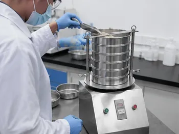
Using a stack of precision sieves on a vibratory sieve shaker—for instance, 500 µm, 100 µm, and 45 µm—you force the sample into disciplined fractions. The specific surface area (SSA), which dictates how a particle adsorbs toxins like pesticides, now becomes a controlled variable. You are no longer guessing. You are comparing like with like.
The Psychology of Reproducibility
There is a quiet terror in the mind of a scientist who cannot reproduce their own results. Variation in particle size distribution (PSD) is one of the most common, yet least acknowledged, causes of inter-laboratory chaos.
By using precision standard test sieves traceable to ASTM or ISO specifications, you replace that terror with confidence. The physical boundary of the mesh ensures that a “100 µm fraction” in Tokyo means the same thing as a “100 µm fraction” in Berlin. Standardization is not bureaucracy; it is scientific sanity.
Enhancing Signal Integrity: When Chemistry Meets a Clean Filter
Fluorescent labeling of microplastics is a common technique. You dye the plastic with Nile Red, then wash off the excess dye. But if the washing is incomplete, unbound dye remains, creating a screaming background fluorescence that drowns out your true particles.
The 45 µm Rinse Protocol
A fine-aperture precision sieve—say, 45 µm—is the unsung hero here. After staining, you dump the suspension onto the sieve. The dye solution drains. You rinse with a solvent while the particles stay trapped. In minutes, you’ve removed the chemical noise.
This applies equally to thermal analysis (pyrolysis-GC/MS) and spectroscopy. When particle size is tightly controlled, you eliminate the variance in thermal lag and mass transfer that skews regression models. The result is a cleaner chromatogram, a sharper spectrum, and a regression coefficient you can actually trust.
The Hidden Traps: What Your Sieve Won’t Tell You
No tool is a panacea. Precision sieves come with their own psychological blind spots that can sabotage a workflow.
The Clogging Nightmare
“Near-size” particles and sticky biofilms can blind a mesh. You assume your 100 µm sieve is letting particles through, but in reality, the effective pore size has shrunk. Smaller microplastics back up and slide over the rim during rinsing. You’ve just lost them. Visual inspection is a lie—a blinding sieve looks identical to a clean one until you measure flow rate.
The Ghost of Samples Past
Cross-contamination is the researcher’s shame. A single microplastic fragment lodged in the transition between mesh and frame can dislodge in a later run. If you don’t meticulously clean your sieves using an ultrasonic bath with a compatible solution, you are measuring memory, not reality. Stainless steel mesh offers durability, but the grain boundaries on cheap wire can harbor residues like a criminal hiding evidence.
Chemical Deterioration
Digestion chemicals are violent. Repeated exposure to concentrated acids or alkalis can slightly enlarge the aperture of a sieve or corrode the wire surface, making it sticky. When the very standard that defines your cut-off begins to drift, your data drifts with it.
This is where equipment quality shifts from a purchasing decision to a scientific obligation. Premium stainless steel test sieves, built with precise tensioning and chemical-resistant frame materials, are the antidote. Combined with automated vibratory sieve shakers from a provider who understands the full sample preparation chain, you mitigate these risks before they become retracted papers.
Your Workflow, Your Rules: A Decision Framework
Not every research goal demands the same approach. Match your sieve strategy to your question.
| If your primary focus is… | Then you must… | The core tool |
|---|---|---|
| Toxicological modeling | Strictly control PSD and SSA for accurate ingestion data | Stack of high-precision sieves (e.g., 500 µm, 100 µm, 45 µm) on a vibratory shaker |
| High-throughput screening | Automate grading to reduce manual solid-liquid separation labor | Air-jet or vibratory sieve shakers with standard meshes |
| Fluorescence quantification | Eliminate unbound dye without losing target particles | Fine-aperture sieve (45 µm) for post-dye rinse phase |
| Thermal or spectroscopic analysis | Remove fines and coarse fragments to normalize responses | Single intermediate mesh (100 µm) to isolate a clean fraction |
An Engineer’s Grace: Why the Sieve Persists
There is a beautiful absurdity in modern science. We use gene sequencers and synchrotrons, yet the most pivotal step in isolating man-made plastic particles from a mussel’s gut is often a disc of woven metal developed from the milling traditions of centuries past.
The sieve does not think. It does not require calibration certificates that cost more than a car. It works because the laws of physics don’t negotiate. An opening of 100 µm is an absolute. In a research field fogged by nanoplastics we cannot see and polymers we misidentify, that absolutism anchors us.
But even an absolute needs a reliable custodian. From the jaw crusher that initially breaks your frozen tissue, to the liquid nitrogen cryogenic grinder that embrittles it, to the precision test sieve that gives you an honest fraction—the chain of custody matters. A complete laboratory sample preparation solution, encompassing milling, sieving, mixing, and pressing, doesn’t just provide instruments. It provides a seamless logic.
When you hold a clean, sized sample in a glass vial, ready for the Raman stage, you feel a rare scientific certainty. That certainty began the moment the liquid passed through the mesh, leaving only what matters behind.
Quick Links
Suggested Products
Laboratory Vibratory Test Sieve Shaker for Precision Particle Size Analysis and Powder Grading Small Laboratory Vibrating Sieve Shaker for Precise Particle Size Analysis Stainless Steel Rotary Vibrating Sieve High Precision Circular Vibratory Separator Industrial Powder Grading Machine Multi Layer Sifting Equipment Stainless Steel Laboratory Vibratory Test Sieve Shaker 三维电磁微量振动筛分仪Further Reading
The Sieve Never Lies: How a Simple Stack of Mesh Became the Lab’s Ultimate Truth-Teller The Grain Size That Held Up Cathedrals: A Sieve Analysis Story The 0.15 mm Question: Why Sieve Grading Is the Quiet Foundation of Reliable Lightweight Mortar The Sieve and the Mind: Why Your Soil Test Results Depend on Replacing Human Fatigue with Engineered Consistency The Quiet Gravity of a Perfect Mix: How Sieves and 3D Tumblers Engineer Certainty in Drug Formulation
PowderPreparation
Last updated on May 14, 2026
Related Products
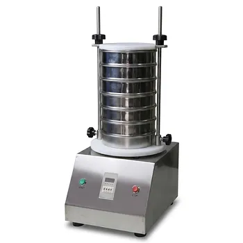
Laboratory Vibratory Test Sieve Shaker for Precision Particle Size Analysis and Powder Grading
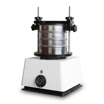
Small Laboratory Vibrating Sieve Shaker for Precise Particle Size Analysis
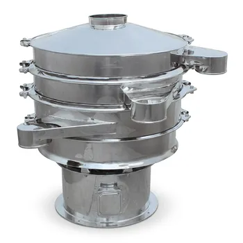
Stainless Steel Rotary Vibrating Sieve High Precision Circular Vibratory Separator Industrial Powder Grading Machine Multi Layer Sifting Equipment
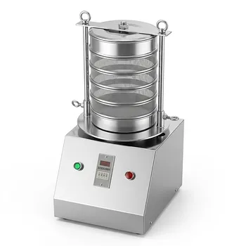
Stainless Steel Laboratory Vibratory Test Sieve Shaker
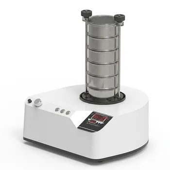
三维电磁微量振动筛分仪
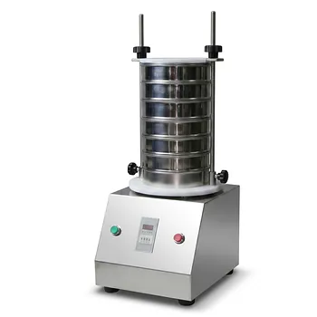
Small Laboratory Vibratory Sieve Shaker for Powder Gradation and Particle Size Analysis
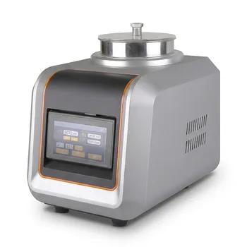
Laboratory Air Jet Sieving Machine for Fine Powder Particle Size Analysis and Deagglomeration
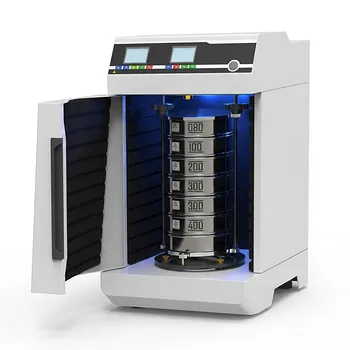
Laboratory Dry and Wet Three Dimensional Vibratory Sieve Shaker for Particle Analysis
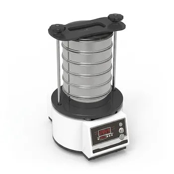
Tapping Oscillating Sieve Shaker for Dry and Wet Particle Size Analysis
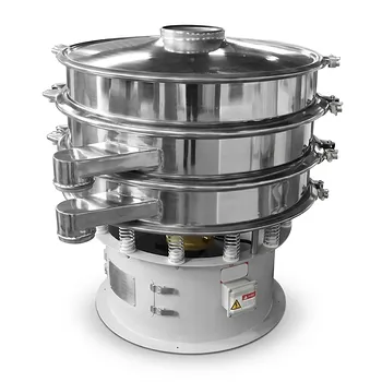
Three Dimensional Rotary Vibrating Sieve
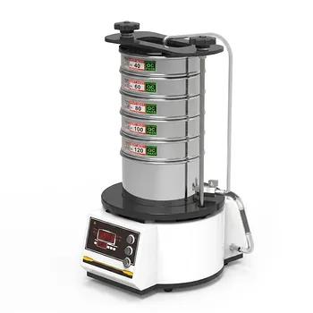
High Frequency Wet Three-Dimensional Vibrating Sieve Shaker for Dry and Wet Particle Size Analysis
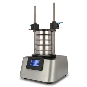
Vibratory Sieve Shaker Electromagnetic 3D Motion Powder Particle Size Analyzer for Dry and Wet Sieving
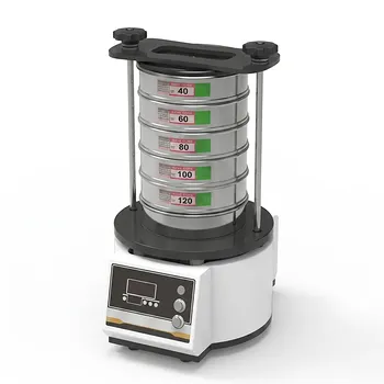
Dry Three Dimensional Vibratory Sieve Shaker
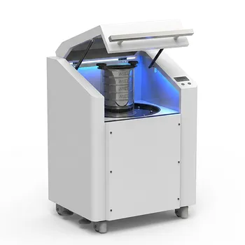
High Frequency Cabinet Type Three-Dimensional Rotary Vibrating Sieve Shaker for Dry Sieving and Particle Classification
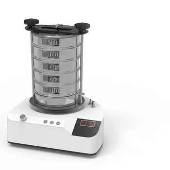
Heavy Duty Dry Three Dimensional Vibratory Sieve Shaker for Particle Separation
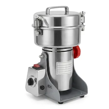
Small High-Speed Laboratory Grinder for Rapid Sample Preparation
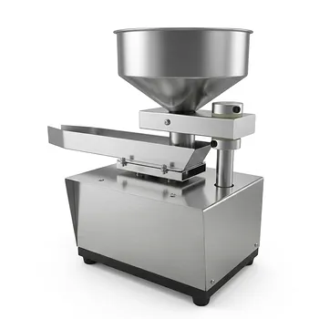
Automatic Vibrating Powder Feeder for Laboratory Material Processing Precision Vibratory Hopper Feeder for Granular and Powder Material Handling Industrial Grade Vibrating Tray Feeder for Consistent Material Sample Preparation
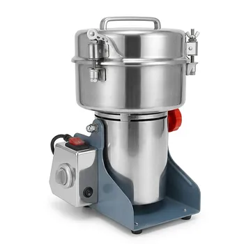
Small High-Speed Grinder for Efficient Laboratory Sample Preparation